
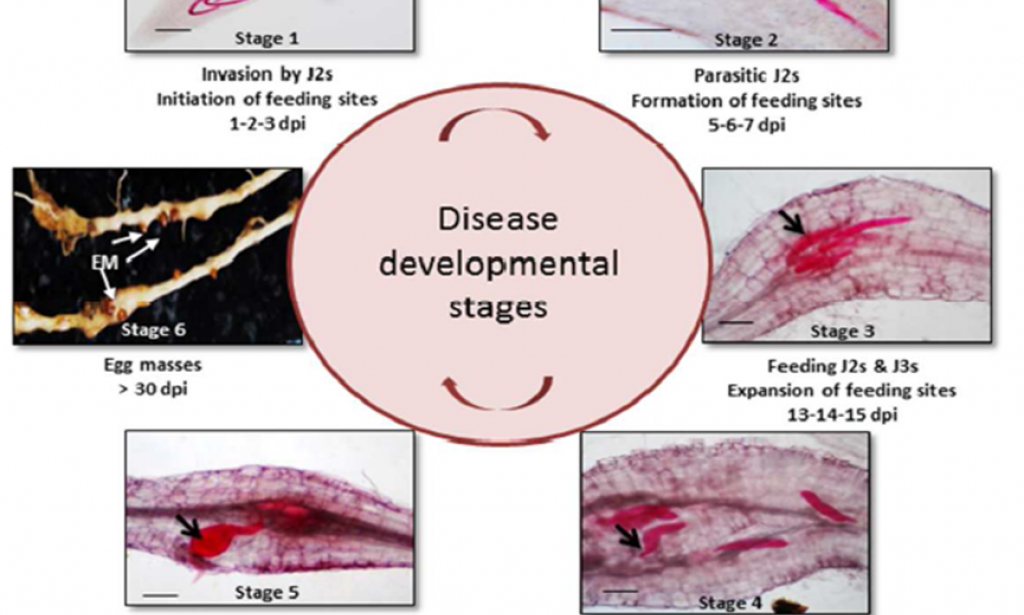
The life cycle of plant-parasitic root-knot nematodes consists of two phases: the pre-parasitic and parasitic phase. Pre-parasitic phase, root-knot nematodes survive in roots of weeds or as eggs in infected roots of previous crops. The parasitic phase started when the eggs hatch the second larva, go through third, fourth larval stages, and then become adult. The duration of the life cycle is almost four weeks but depend on temperature and the host species. During parasitic phase, the second stage of juveniles (J2) hatches from the eggs in response to host-plant root exudates and invade the root just behind the apex, preferentially in the differentiation and elongation zone (Abad, et al., 2003).
Then, migrated inter-cellularly between the cortical cells down towards the root tip and when reach vascular cylinder, the nematode make a U-turn in order to enter into the cylinder. There, where RKN often involving complex morphological and physiological alterations of host cell into specialized cell called giant cells(Gheysen and Mitchum, 2011). Fully differentiated giant cells are essential to complete the life cycle of RKN for duration of four weeks.During these developmental periods, the nematodes feed alternatively from the different giant cells. After three molting they grow entirely into female adults, which reproduce by parthenogenesis (most species) and deposit their eggs in gelatinous egg sacs (Williamson and Gleason, 2003).
Root-knot nematodes reproduce by three types of reproduction, amphimixis, facultative meiotic parthenogenesis and obligate mitotic parthenogenesis. Only seven of 37 species of Meloidogyne studied to date are amphimictic. Like many soil nematodes, most Meloidogyne spp. are parthenogenesis. Some are facultative meiotic parthenogenesis, and several of the most widespread and economically important species are obligate mitotic parthenogenesis. Populations of the same Meloidogyne species may differ in mode of reproduction; for example, 29 of 32 populations of M. hapla reproduced by facultative meiotic parthenogenesis, the others were mitotic parthenogenesis (Triantaphyllou, 1966). Males migrate out of the plant (Fig. 7.1A) only a few species, e.g. M. carolinensis, M. microtyla and M. pini, reproduce by amphimixis, with the obligatory fusion of a male and female gamete. After the development of the pear-shaped female, eggs are released on the root surface in a protective gelatinous matrix. Embryogenesis within the egg is followed by the first molt, leading to the J2.

You must be logged in to post a comment.